| Fossils are critical for understanding the evolutionary diversification, turnover, and morphological disparification of extant lineages. While fossils cannot be sequenced, phenome-scale data may be generated using micro-computed tomography (μ-CT), thus revealing hidden structures and
internal anatomy, when preserved. Here, we adduce the male caste of a new fossil ant species from Miocene Ethiopian amber that resembles members of the Aneuretinae, matching the operational definition of the subfamily. Through the use of synchrotron radiation for μ-CT, we critically test the aneuretine-identity hypothesis. Our results indicate that the new fossils do not belong to the Aneuretinae, but rather the Ponerini (Ponerinae). Informed by recent phylogenomic studies, we were able to place the fossils close to the extant genus Cryptopone based on logical character analysis, with the two uniquely sharing absence of the subpetiolar process among all ponerine genera. Consequently, we: (1) revise the male-based key to the global ant subfamilies; (2) revise the definitions of Aneuretinae, Ponerinae, Platythyreini, and Ponerini; (3) discuss the evolution of ant mandibles; and (4) describe the fossils as †Desyopone hereon gen. et sp. nov. Our study highlights the value of males
for ant systematics and the tremendous potential of phenomic imaging technologies for the study of ant evolution Insects 2022, 13, 796. https://doi.org/10.3390/insects13090796 Boudinot, B.E.; Richter, A.K.; Hammel, J.U.; Szwedo, J.; Bojarski, B.; Perrichot, V. | 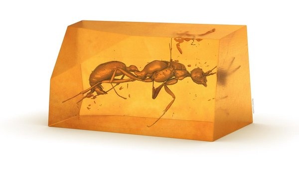 |